sphenopalatine sinus horse
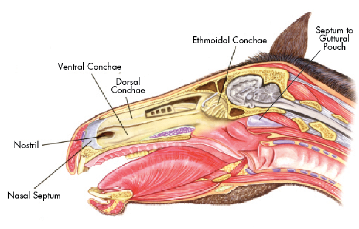
Due to the size and complex anatomy these disease processes can sometimes be present for weeks or months before clinical signs such as nasal discharge or facial swelling are evident. Inspissated thickened pus accumulated in this horses sinuses especially in the ventral conchal sinus.

Learn More With This Infectious Disease Article By R Wayne Waguespack And Jennifer Taintor
Sphenopalatine sinus anatomy was described and compared between cadaver specimens across the imaging modalities.

. In the horse the sphenoid and palatine sinus compartments communicate and are hence known as the sphenopalatine sinus. The horses head has uniquely adapted itself and developed six pairs of paranasal sinusesthe frontal sphenopalatine and maxillary sinuses and the dorsal middle and ventral conchal sinuses. In the horse the maxillary sinus has caudal and rostral parts which together occupy a large part of the upper jaw.
1 Treatment of chronic equine. The borders of the sphenopalatine sinus were not identifiable on plain. Cadaver study and client-owned 20-year-old Warmblood gelding.
The neuro-opthalmological picture of sphenopalatine sinus syndrome has been theorized in horses on the basis of the complex anatomy of that sinus cavity. Sphenopalatine sinus syndrome in a horse. The sphenoidal and palatine sinuses communicated in most horses.
The anatomy of the sphenopalatine sinus was variable. The sphenoidal and palatine sinuses communicated in most horses. In terms of management lavage flushing out is.
In such cases what could accurately be termed the combined sphenopalatine sinuses usually drained directly into the caudal maxillary sinuses. Disorders of the equine sphenopalatine sinus including empyema and neoplasia have been reported to cause damage to cranial nerves II and V. In such cases what could accurately be termed the combined sphenopalatine sinuses usually drained directly into the caudal maxillary sinuses.
General terms Splanchnology Respiratory system Paranasal sinuses Sphenopalatine sinus. However the clinical anatomy of these sinuses is not well described in horses. ArticleBertuglia2006SphenopalatineSS titleSphenopalatine sinus syndrome in a horse authorAndrea Bertuglia and Antonella Rampazzo and A Brignolo and Antonio Dangelo journalIppologia year2006 volume17 pages13-16.
The maxillary sinus is the largest paranasal sinus and is divided into two parts rostral and caudal by a thin septum. In addition the horse has sphenopalatine and ethmoidal sinuses which are of a lesser clinical importance than the frontal caudal maxillary and rostral maxillary sinuses on each side of the. Additionally in 5 out of 16 cases some compartments of the sphenoidal sinus also drained into the ethmoidal sinus.
To describe a novel standing trans-nasal endoscopic guided CO 2 laser fenestration approach to access the sphenopalatine sinus SPS in the horse. A few anecdotal cases had been reported in. Request PDF Sphenopalatine sinus syndrome in a horse A 14-years-old Arabian mare presented for clinical consultation with a 2-months history of chronic nasal discharge and recurrent epistaxis.
Bacterial meningitis in adult horses has been described as a rare life-threatening complication of dental disease periocular pathology sinusitis andor sinus surgery 12Clinical signs include cervical pain or stiffness with reluctance to flex the neck fever lethargy and muscle fasciculations The most common neurologic sign includes ataxia that. To get spatial sense of the equine specific communication ways between the nasal cavity and the paranasal sinuses heads of 19 horses aged 2 to 26 years were analyzed using three. Additionally in 5 out of 16 cases some compartments of the sphenoidal sinus also drained into the ethmoidal sinus.
However the clinical anatomy of these sinuses is not well described in horses. In human medicine it is described that obstruction of the sinonasal communication plays a major role in the development of sinusitis. Sinusitis is a common disease in the horse.
Medical records January 2004-2014 of cases diagnosed with sphenopalatine sinus disease were reviewed. Conclusion Transnasal endoscopically-guided ventral surgical access to the sphenopalatine sinus is possible in horses and may improve access in horses with disease extending caudally beyond the palatine portion. These cavaties also extend around above and below the horses eyes and end around the facial crest 1.
This sinus lies under the ethmoidal labrynth. The frontal sinus has 3 chambers which drain separately into the nasal cavity. Paranasal sinus disorders include primary or secondary sinusitis progressive ethmoid hematoma sinus cysts and less commonly neoplasia.
Premolars and molar tooth roots are also found in the simus. Sphenopalatine sinus - Sinus sphenopalatinus. The sinuses are six air-filled cavities within the head of the horse - the frontal sphenopalatine and maxillary sinuses and the dorsal middle and ventral conchal sinuses.
Reasons for performing study. The sphenopalatine sinus drains via the caudal maxillary sinus with which is communicates freely over the infraorbital canal. Radiographic computed tomographic and surgical anatomy of the equine sphenopalatine sinus in normal and diseased horses Author.
The horse recovered well from surgery and although it has not regained vision as of 65 years postoperatively the disease has not progressed.
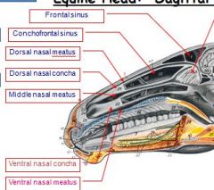
Anatomy Ii Head Equine Flashcards Cram Com

Pdf Nasal And Paranasal Sinuses Of The Donkey Gross Anatomy And Computed Tomography

Equine Paranasal Sinus Disease Questions Facial Swelling And Nasal Discharge Flashcards Quizlet

Frontal Sinus An Overview Sciencedirect Topics
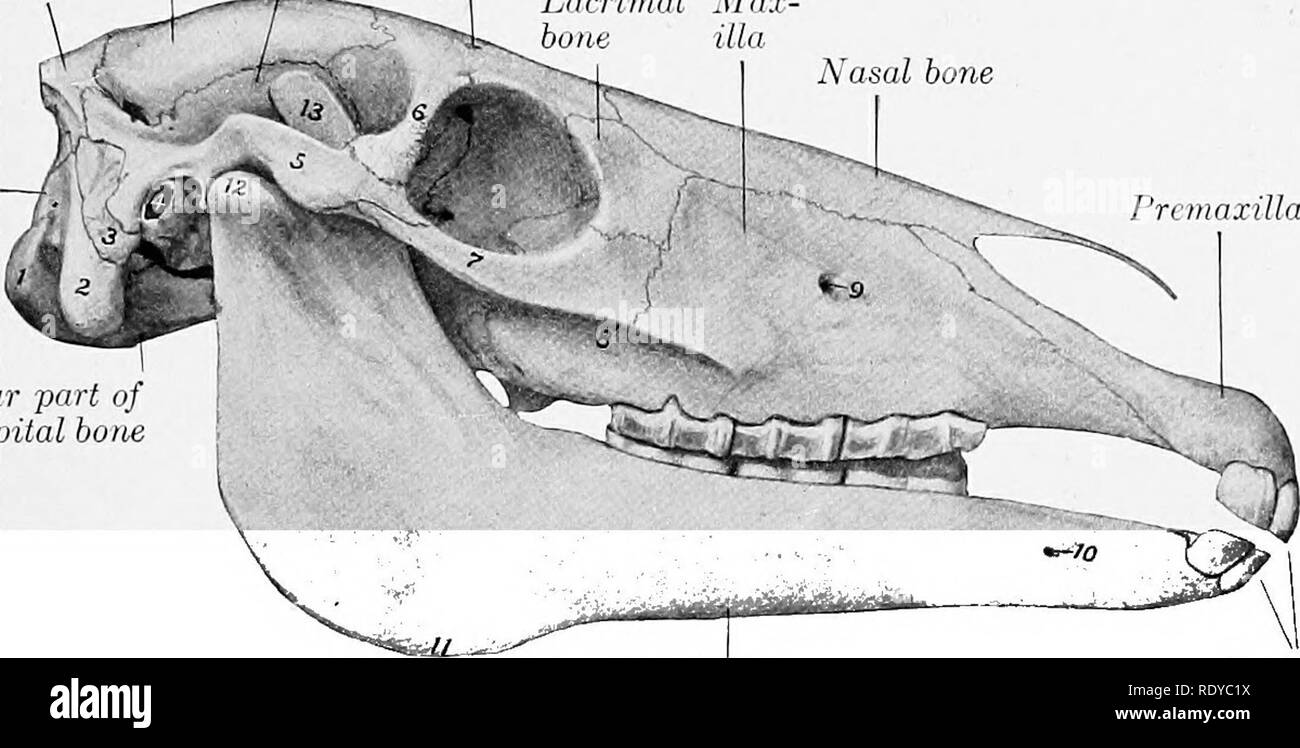
The Anatomy Of The Domestic Animals Veterinary Anatomy The Paranasal Sinuses 85 Superior Maxillary Sinus Is Also Crossed By The Infraorbital Canal Over Which It Opens Freely Into The Sphenopalatine

The Anatomy Of The Domestic Animals Veterinary Anatomy The Paranasal Sinuses 85 Superior Maxillary Sinus Is Also Crossed By The Infraorbital Canal Over Which It Opens Freely Into The Sphenopalatine Sinus

3d Models Of The Skull Semitransparent And Paranasal Sinuses Download Scientific Diagram

Equine Head Special Structures Of The Paranasal Sinuses Flashcards Quizlet

Equine Cardiovascular Respiratory Flashcards Quizlet

Equine Nasal Discharge Flashcards Quizlet

Sagittal Sections A B 12 Years C 8 Years Of The Head Of The Donkeys Download Scientific Diagram
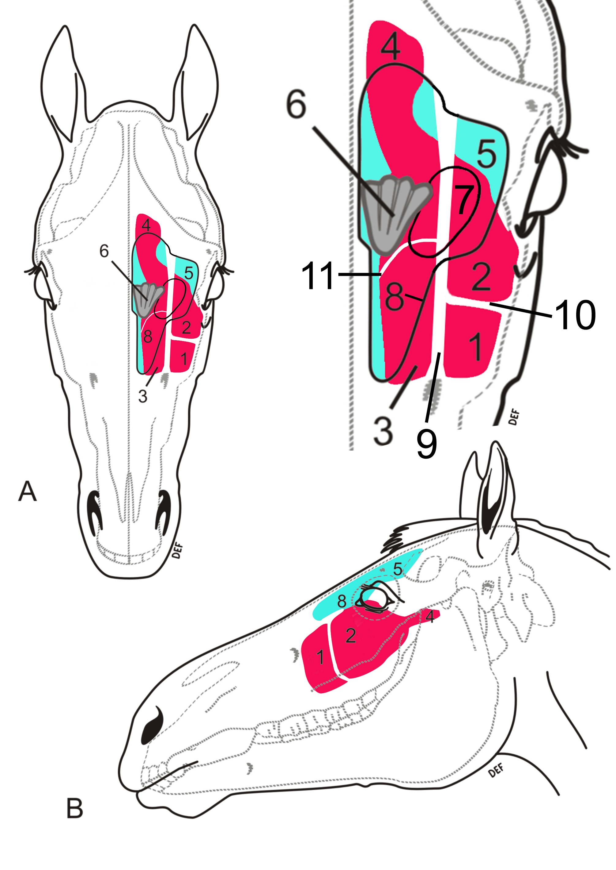
Equine Sinus Conditions Large Animal Hospital College Of Veterinary Medicine University Of Florida

Horses With Facial Swelling Nasal Discharge Flashcards Quizlet
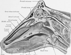
Nasal Discharge Large Animal Surgery Supplemental Notes

Direct Approach To The Nasal Cavity Through A Bone Flap For The Treatment Of A Large Nasal Cyst Bolz 2017 Equine Veterinary Education Wiley Online Library

The Anatomy Of The Domestic Animals Veterinary Anatomy The Paranasal Sinuses 85 Superior Maxillary Sinus Is Also Crossed Bj The Infraorbital Canal Over Which It Opens Freeh Into The Sphenopalatine Sinus
Equine Sinus Disease A Hidden Danger
0 Response to "sphenopalatine sinus horse"
Post a Comment